Five Steps To Prevent Tendon Injuries In Your Horse

Tendon injuries can mean lengthy time off, stall rest, etc. for your horse, so all of us want to do everything possible to prevent them from occurring. Here are five steps on how to prevent them:

1. Buy a horse with the best conformation possible. We all realize that there is no perfectly conformed horse and there are definitely cases where you have to give up a little of the conformation for a horse with a good brain or that is really good at its job, but ideally you buy as good a conformation as possible, especially for your particular discipline. Certain conformations are more prone to various injuries.  Horses that are very straight through their hind end are more prone to hock and high suspensory issues. Horses with long pasterns and underrun heels are more prone to suspensory issues in the front limbs and/or various other tendon issues. Horses that toe in put a lot of strain on the ligaments on the outside of their limbs. If you can buy a horse that minimizes these conformational faults, you are setting yourself up better for success.

2. Shoeing is key. Have a good farrier and keep your horse on a regular schedule. Again, horses that have conformational faults will put more strain on their ligaments at the end of a shoeing cycle when toes are long and heels are underrun due to greater leverage on these areas. So keeping them on a regular cycle is key. Also certain things can be done with trimming and shoeing to minimize stresses on various areas. So find a farrier you trust. Consult with your vet. And have the two of them work together.

3. Fitness. We all know what it’s like to be a weekend warrior and not exercise all week and then go out and overdo it on the weekend. Horses are no different. So don’t go out and expect to do a 50 mile endurance ride after not riding your horse for several weeks and expect him not to be sore. The more fit your horse is the less likely that he is to fatigue while you’re riding him. Injuries often happen when your horse is fatigued and not taking care of their body as well as they could. These tendons and ligaments also get tired and are more prone to injury at this time.

4. Terrain: Ride your horse on different types of terrain. Always riding on the same terrain makes tendons and ligaments accustomed to only that type of terrain. It makes sense to ride them on different types of ground.  Ride them on hard ground. Ride them on grass. Ride them in an arena. Ride them carefully over uneven ground. All these things will strengthen your horse’s ligaments and make them less prone to tearing.

5. Cross train your horse. This means ride them in an arena. Take them out on trail rides. Do trot sets. Ride them long and low. Ride them more collected. This will help strengthen your horse’s core and strengthen their hind end.  Also getting them to use their core will help strengthen their back and horses can get sore backs just like we can.

Equine Shockwave: What Is It & What Does It Do?

Extracorporeal shockwave therapy, or shockwave, is a non-invasive technique using sound waves to stimulate healing in wounds, ligaments, tendons, and bony structures.  It is thought to increase blood flow circulation and growth of blood vessels to the treated area.  By doing this, natural healing factors are then able to get to the affected area and decrease inflammation and help stimulate healing.  In addition to stimulating healing by this manner, it also provides some mild, short term pain relief.

Shockwave is commonly used in cases of suspensory ligament disease, tendon disease, back pain, bone bruising, painful splints, bucked shins, and navicular syndrome.  Horses are often sedated for this procedure and usually it lasts no longer than 30 minutes.  The area that is receiving treatment is cleaned and a gel applied to help the sound waves get transmitted from the transducer probe.  The transducer is then applied to the area and emits high-energy sound waves.  The intensity of the shocks and the proper number of impulses that the horse is treated with depends on the area affected.  Typically the deeper the tissue the affected, the greater the number of impulses and energy required.

Many different types of shockwave exist and not all are created equal.  Blue Ridge Equine Clinic uses a system which generates an electrohydraulic shockwave which allows this system to penetrate deeper than many other systems.  Also this system has multiple different probes that allow it to treat different focal areas depending on the condition being treated while generating a true shockwave at all energy settings.  Appropriate focal area, along with effective shockwave penetration together create the most effective shockwave for our medical purposes.

Currently most racing jurisdictions prohibit the use of shockwave within 5-7 days of racing, and the FEI prohibits the use of shockwave within 5 days of competition. The USEF recommends a three day withdrawal period before competition, although it may be administered by a veterinarian no closer than 12 hours before competition and only in the application to the back and dorsal pelvis areas.  It is due to the pain relief effect of shockwave that these time periods have been chosen.

In addition to proper rest and rehab, shockwave can be a very useful adjunct therapy in many cases of lameness and orthopedic disease.  In many cases insurance will allow up to three treatments of shockwave if recommended by the veterinarian.  Please contact your veterinarian if you have any questions regarding shockwave.

Second Chance For Horse With Severe Laminitis

Clara Louise had been battling chronic laminitis for months, but suddenly things went from bad to worse. At 13 years old, the TB/Welsh cross mare was now so severely lame that she was reluctant to move. She had been on medical treatment for the chronic laminitis, including Prascend® to treat her Pituitary Pars Intermedia Dysfunction (PPID, also known as Equine Cushing’s Syndrome). When Dr. Reynolds Cowles examined her, her front feet were acutely painful, and radiographs showed severe rotation.

Anti-inflammatory medications were added and special support shoes with a wedge to take pressure off of the deep digital flexor tendon were taped on. The deep digital flexor attaches on the underside of the coffin bone, and its pull contributes to rotation of the bone when the attachments to  the hoof wall are weakened by inflammation. The shoes helped some, but Clara Louise was still in a lot of pain. It was decided that surgery to cut the deep digital flexor was needed to relieve the pull on the coffin bone. Clara Louise’s owner, Kathy Buhrer, brought Clara Louise to Blue Ridge Equine Clinic for surgery and corrective shoeing.

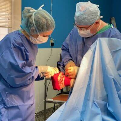
By the time Clara Louise arrived at Blue Ridge Equine Clinic, she was extremely painful and could barely walk.  Dr. Marcos Santos blocked her lower legs and feet so that farrier John Kern, with the assistance of  Marcus Wise, and Dr. Santos could work on her without causing her pain. Under the direction of Dr. Santos, the farrier carefully trimmed her feet and glued on shoes with extra support behind the foot and underneath the frog.  Dr. Santos then performed a standing tenotomy, a surgery where the deep digital flexor tendon is cut, relieving the pull on the coffin bone.

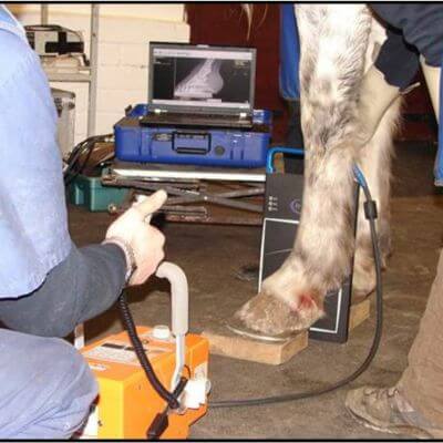
Radiographs taken at the time of surgery demonstrate how much the expert trimming and surgery helped to re-align the coffin bone in the hoof capsule. This video shows her the day after surgery, again a few days later, and on day five post surgery.

Clara Louise was maintained on anti-inflammatory medication, pain management, and medications to protect her digestive tract. She made very good progress, and soon returned to a farm near the clinic. Dr. Santos visited her regularly to change her bandages and carefully monitor her progress. This video shows her after her second shoeing cycle, post surgery. She has returned home, and is continuing to feel better all the time. Recent radiographs show that Clara Louise’s feet are now essentially normal. Her story isn’t over, but everyone on Clara Louise’s team has been thrilled and grateful for her progress; certainly not every horse or pony with severe laminitis has so happy an outcome.  Her story shows that with hard work, know-how, commitment, and some good fortune, some severely affected patients can make a recovery.


Latest update from Clara’s owners: “Look at this beautiful, happy pony!!!! Such a difference from Dec 2. I update Dr Santos weekly, will you share with John the farrier? Let him know this recent shoeing seemed to really make a major difference, I don’t know if they changed anything  but she is (fingers still crossed) doing soooooo well.”

Ristocrat’s Return: Making The Most Of Time Off

There it was, the news that horse owners dread to hear: the young, beautiful, talented warmblood that she bred had a suspensory tear. All the momentum of his promising career in dressage seemed to have ground to a halt, and now a new goal loomed. Roberta Falk was determined to pursue the best course of treatment for Ristocrat to put him back on the road to success.

The diagnosis was made through performing regional anesthesia (selectively blocking nerves in the lower leg to determine where the pain is) and ultrasound. The ultrasound examination also gave the information that Ristocrat’s injury was likely to benefit from regenerative treatment. It was decided to treat the tear with stem cells. Appointments were made, stem cells were harvested and processed, and then injected into the injured areas using ultrasound to guide the needles.

Regardless of other interventions, time and rest and controlled exercise (emphasis on controlled) are the basis of healing any soft tissue injury. Most horses spend 8-9 months, sometimes more, in a program of slowly increasing exercise before they are back to full work and allowed unrestricted turn out. Each animal is an individual, and every injury heals at its own speed, making the timing of changes to the program difficult. This timing is also key to keeping the progress going as efficiently as possible while avoiding further injury. The use of ultrasound to monitor the healing of these structures has vastly improved our ability to successfully guide rehabilitation in these injuries, improving outcomes from up to 50% success to up to 80% success.

Ristocrat had some challenges up his sleeve, though. Shortly after he returned home after his regenerative medicine therapy, his leg swelled up and was hot. Ultrasound of the leg confirmed that his body was having an exaggerated response to the biological therapies. Cold hosing, wrapping, and a few days of bute got things quieted down again. Fortunately, he was happy in his stall and enjoyed his hand walking, and when he was checked again the next time, significant healing was seen on ultrasound, and he graduated to the next level of exercise.

As he was asked incrementally to do more work, his muscles and joints were kept loose and limber with chiropractic adjustments. Little by little, more work was added at a pace dictated by the pace of his healing. He returned to full work, and was working well. He started showing, and he was doing well. After a short vacation, however, it was noted that he would skip off of that front leg at the trot in tight turns. After cantering, his gait was normal. Ultrasound examination of the suspensory showed some scar tissue at the old injury site. Shock wave was used along with exercise to break down the dense scar so that the ligament could stretch freely. The skipping has not returned.

Without ultrasound, determining the pacing of rehabilitation is difficult at best. Horses that heal more slowly need to progress through their rehabilitation program at a slower pace, and this cannot be determined by the horse’s soundness alone. Not all horses can be on the same schedule. Also, knowing the cause of a gait problem (injury vs. scar tissue) allows the correct course of action (rest vs exercise) to be determined. Ultrasound has helped Ristocrat get back to work efficiently and safely. Once again, the future is bright for this lovely sales prospect and his team.

Acupuncture To Treat Nerve Damage: How Caddie Got Back On His Feet

The practice of veterinary acupuncture is rooted in the tenets of Traditional Chinese Medicine and the principles of neuro-humoral responses as explained by modern Western Medicine. Acupuncture is most commonly used for relief of musculoskeletal and neurologic pain, reducing anxiety, and improving the function of damaged nerves. Electroacupuncture is a technique that transmits low levels of electricity into the tissues, and can be very useful for relief of chronic pain and for re-establishing the flow of impulses in nerves that have been damaged by trauma or infection. An example of how electroacupuncture can be helpful is Caddie Johnson.

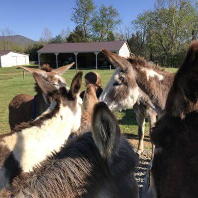
Caddie Johnson is a donkey that had not gotten many breaks in his young life. He became ill when very young, requiring veterinary treatment. He seemed to recover, but his owners Dave and Barbara noticed an abnormality in his gait. At first, it seemed to come and go, and shift from one front leg to the other, which made finding the cause very difficult. He also seemed fatigued and listless. When Caddie started knuckling over in his fetlocks, it seemed that the problem was in his neck, and not in his legs. Ultimately, Caddie was diagnosed with an infection in his neck bones that was pressing on the nerves that ran down his front legs. This is a relatively rare and very serious condition that is very challenging to treat. The infection responded well to long term antibiotic use, and although Caddie was feeling much better overall, he was still knuckling on the front legs, especially the left front. He was given several months’ time to heal, but the nerves were still not working.

Anyone who knows donkeys will understand that it was with some trepidation that I approached placing needles in the legs of this yearling Jack, but with some reassurance to Caddie from his loved ones, we prevailed, and surprisingly, Caddie stood like a champ when the electrodes were attached and the current applied. If anything, he got a little sleepy. The plan was to repeat the treatment every 1-2 weeks and see how things went. After the first treatment, the Johnsons reported that Caddie’s gait was better and that he seemed to have more energy. The following two treatments were much more challenging than the first; Caddie was very rambunctious and unwilling to stand still. Despite his opinions on the matter, he did get treated, and after 3 treatments, Caddie was back to his old self, playing in his field and full of spunk.

Diagnosing Lameness In Horses: New Tool For Blue Ridge Equine Clinic

Blue Ridge Equine Clinic is excited to announce that it has recently purchased a new piece of equipment that can help us more accurately diagnose subtle lameness and performance problems.

The Lameness Locator is a handheld computer system that helps veterinarians objectively identify lameness using noninvasive inertial sensors.  Motion sensors are placed on the horse’s poll, pastern, and pelvis and are wirelessly tracked by a portable computer.  The equipment measures vertical acceleration of the lameness locator2torso to determine asymmetries in head and pelvic position between left and right halves of stride.  This data is transmitted instantly to the portable computer which contains a computer program that uses algorithms to translate this data into a graphical representation of the lameness.  These algorithms were developed by years of extensive motion analysis research.  The data from the sensors can show the location (limb(s)) of asymmetry in the gait, the severity of the lameness, and at what stance in the gait the lameness appears.

The Lameness Locator sensors sample data faster than the human eye, allowing us to detect things that were previously undetectable. It is especially helpful in horses with very mild lameness or history of “poor performance” that may be difficult to see with the naked eye.  It is also helpful in horses with multiple limb lameness, and may help the veterinarian determine which lameness is the primary lameness and also at what stance in the gait the lameness appears. This may give insight as to from where in the limb the lameness is coming. The equipment makes lameness exams more accurate by providing a scientific measure of the improvement in gait following a nerve block. The Lameness Locator could potentially improve prognosis by allowing veterinarians to diagnose and initiate treatment earlier in the course of disease or injury. We are excited to offer this technology as yet another tool to help maximize performance in our sporthorses, and to help aid our diagnosis in difficult lameness cases.